Thursday 30 December 2010

Brain MRI Indicates Link Between Amygdala Volume And Social Network.

In a recent study, brain MRI scans showed that the more varied social network an individual has, the bigger his/her amygdala will be. The study was carried out by researchers from Massachusetts General Hospital (MGH) in the US. The findings appear in the 26 December advance online issue of Nature Neuroscience.
The amygdala consists of 2 symmetrically placed small almond shaped structures located deep into the temporal lobe. It is connected to various brain structures and amygdala is involved in a wide range of behavioral functions.  "We considered a single primate species, humans, and found that the amygdala volume positively correlated with the size and complexity of social networks in adult humans. They found the link was just as strong when they adjusted for age (older people have on average smaller amygdala volumes than younger people) and when they analyzed left and right amygdalas separately, indicating no lateralization of the effect" said Dr. Lisa Feldman Barrett, the study co-leader, from MGH's Psychiatric Neuroimaging Research Program and a Distinguished Professor of Psychology at Northeastern University.

To read the rest of this news, please visit:

Brain MRI Indicates Link Between Amygdala Volume And Social Network.

New Screening Tools Offer New Chances For Radiologists

A new regulation has been launched under the Patient Protection and Affordable Care Act affecting practices under the in-office ancillary exception of the Stark law. Referring doctors who offer in-office screening services must offer their patients with a list of other options in their region.


According to this new rule, Thomas W. Greeson, a partner at the Falls Church, Va., office of healthcare group Reed Smith LLP mentioned that most diagnostic imaging centers and radiology groups will not be affected directly, however, he reported that some radiology groups are going to be subjected to the requirement for instance interventional radiologists who are working as treating doctors.

To read the rest of the article, please visit:
New Screening Tools Offer New Chances For Radiologists

CT Scans Can Help In Detecting Lung Cancer In High Risk Populations.

Digital Radiography newsResearchers at the University of Cincinnati reported that CT scans can be more effective in cases of high risk lung cancer, if the scans were followed by clinical routine with a multidisciplinary care team.  


These results have been reached after a clinical trial conducted on a high-risk local population with rates of histoplasmosis, a fungal infection that enters the body through the lungs and increases the likelihood of lung nodules, three times higher than the national average, performed by Dr. Sandra Starnes, director of thoracic surgery at the University of Cincinnati (UC) College of Medicine and a surgeon with UC Health.
In order to read the rest of the article please visit:
CT Scans Can Help In Detecting Lung Cancer In High Risk Populations.

Michelson Diagnostics Chosen To Participate In Future Health Mission 2011

Between more than 70 applicants, Michelson Diagnostics Ltd, the Orpington, UK-based developer and manufacturer of laser scanners for use in non-melanoma skin cancer diagnosis & treatment is chosen to participate in the Future Health Mission 2011 to the USA. The event is taking place in San Francisco along with the annual J.P. Morgan Healthcare Investing Conference in early January.

Regarding to the mission organizers, the Future Health Mission 2011 may provide the businessmen responsible for these 20 growing healthcare technology companies with the opportunity to connect with key people from the US's top technology businesses. Those companies which are the most successful in the UK will manage a conference to meet the media from all over the world as well as investors and potential partners.

For more info. please visit:

Michelson Diagnostics Chosen To Participate In Future Health Mission 2011

Wednesday 29 December 2010

New Progress With The Cooperation Between Calypso Medical Technologies And Elekta

A master development agreement is announced by both Calypso Medical Technologies, Inc., a developer of real-time localization technology used for the precise tracking of tumors, and Elekta, a leading manufacturer of medical devices and software for treating cancer. This cooperation aims to join the developed products integrating the Calypso® System with Elekta's radiotherapy treatment technologies.


Edward Vertatschitsch, Ph.D., President and Chief Executive Officer of Calypso Medical said "The combination of the Calypso System into Elekta's world-class radiotherapy technologies could help to improve innovative treatment modalities used to heal prostate cancer along with more problematic radiation therapy targets, like the pancreas and lung". He added "Some of the desired goals are to connect the linear accelerator and the Calypso System for patient positioning during radiation delivery. This connectivity automates the process of re-positioning the treatment couch when the tumor moves out of a prescribed motion threshold".

For more info. please visit:


New Progress With The Cooperation Between Calypso Medical Technologies And Elekta

Photon-based Radiotherapy Offers Better Results Against Spinal Cord Tumors, Study.

According to a new study, photon-based radiotherapy can be more effective than proton beam while treating spinal cord tumors. The study also showed that photon radiotherapy offers improved survival on long-term. The findings of this study appear in a recent issue of the International Journal of Radiation Oncology, Biology, Physics.
The study is the first to review the long-term results of spinal-cord tumor patients when treated by more modern radiotherapy procedures. According to researchers, Gilomas are the most common spinal cord tumors; they affect around 17,000 US citizens every year, causing death to 13,000 of them.
Dr. Arnab Chakravarti, chair and professor of Radiation Oncology and co-director of the Brain Tumor Program at The Ohio State Comprehensive Cancer Center — Arthur G. James Cancer Hospital and Richard J. Solove Research Institute, said “Our findings need to be verified in a larger number of patients, but they suggest that individuals younger than age 54, those with ependymomas and those treated with photon-based therapy versus proton-beam treatment have better overall survival,”

To read the rest of this news, please visit:


Photon-based Radiotherapy Offers Better Results Against Spinal Cord Tumors, Study.

Ultherapy, An Ultrasound Device Used For Non-Surgical Cosmetic Procedures.

The Laser & Rejuvenation Center of Gold Skin Care has announced the first of its kind in the Middle Tennessee area, Ultherapy™, a unique ultrasound device which received the FDA clearance for lifting the brow. It can also be used to lift and lighten the lower face and neck areas above the thyroid cartilage.


While drawing on the same reliable ultrasound principles that doctors and patients have trusted over years, the Ulthera System is considered a pioneer technology because it utilizes ultrasound energy on the face and neck to lift and tighten the skin in a short, 45-minute procedure. Ultherapy™ has been called the non-surgical facelift procedure, and can be done safely and effectively in the office.

To read the rest of the article, please visit:
Ultherapy, An Ultrasound Device Used For Non-Surgical Cosmetic Procedures.

A New Method For Reducing The Possibility Of Future Osteoporotic Fractures.


digital radiography news
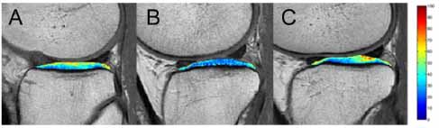
According to the results of the study that was presented at the 2010 congress of the Radiological Society of North America (RSNA) annual meeting in Chicago (IL, USA). Swedish researchers have launched a method by analyzing historic radiology pictures for easing the burden of future osteoporotic fractures.
A study of 8,257 patients at the Karolinska Institute (Stockholm, Sweden) used Sectra’s (Linköping, Sweden) patented digital X-ray radiogrammetry (DXR) way in order to determine patients that suffered from a hip fracture. Through structured osteoporosis prevention program millions of Euros can be protected.
In order to read the rest of the article please visit:

A New Method For Reducing The Possibility Of Future Osteoporotic Fractures.

Tuesday 28 December 2010

SHAPE Starts Registration For Its Carotid IMT Certification Workshop.

SAHPE, The Society for Heart Attack Prevention and Eradication announced that the registration for SHAPE Carotid IMT Certification Workshop has begun. The event will take place on the 15th of January 2011 at Trevisio Restaurant and Conference Center in the Texas Medical Center in Houston. SHAPE is a non-profit organization aiming to improve early diagnosis and preventive measures in order to decrease the risks of cardiac attacks. The workshop will be attended by primary care physicians, cardiologists and radiologists.
The event will take place as a part of the SHAPE Provider Certification initiative, which is concentrating on following the best procedures to decrease the incidence of acute coronary events and mortalities from coronary artery disease. During the workshop, attendees are going to learn evaluate patients’ eligibility to undergo Carotid artery intima-media thickness (IMT) screening, a procedure that is carried out to identify hidden plaques while developing in coronary and carotid arteries. Attendees will also learn to how to use Carotid IMT in a primary care facility.

To read the rest of this news, please visit:

SHAPE Starts Registration For Its Carotid IMT Certification Workshop.

One In Six Physicians In Washington Lease Or Own Medical Screening Tools, Study

According to a study, 1 in 6 doctors in 2008 mentioned that her or his practice leased or owned improved screening tools. Doctor practice leasing or ownership of a wide group of medical tools ranging from tools for laboratory examining to strict proceedings, is quite common.


The Center for Studying Health System change (HSC) launched the report which was supported by the Robert Wood Johnson Foundation. More referrals would be formed by doctors with or other financial interests in medical facilities than medically necessary. The federal physician self-referral law, identified as the Stark Act since the mid-1990s, has forbidden doctors from self-referring Medicare and Medicaid patients to a specific health services, ranging from clinical laboratory services to improved screening services.

To read the rest of the article, please visit:
One In Six Physicians In Washington Lease Or Own Medical Screening Tools, Study

Radio Frequency Is Used In Florida Hospitals To Detect Missed Surgical Objects Inside Patients.

Healthcare IT news
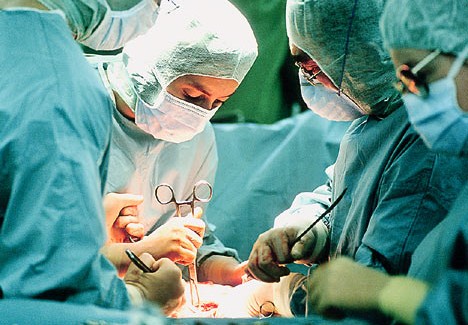
Radio frequency (RF) detection technology is used for the first time in Good Samaritan Medical Center and West Boca Medical Center in the operation rooms to prevent missing any foreign items my mistake inside the patient during the surgery.
Good Samaritan and West Boca are conducting the RF Surgical Detection System from Bellevue, Wash.-based RF Surgical Systems as an assistant to the standard practice of manual counting to improve the patient safety in all conditions at the surgical operations.
In order to read the rest of the article please visit:
Radio Frequency Is Used In Florida Hospitals To Detect Missed Surgical Objects Inside Patients

Monday 27 December 2010

50% Of Females Over 40 In US Get Annual Mammography Scans, Study.

According to a new study, 50% only of the females over the age of 40 in the US undergo annual mammography scans for early detection of breast cancer, even if their health insurance is covering the procedure. The study was discussed at the annual San Antonio Breast Cancer Symposium, which took place earlier this month.
The research team reviewed database of around 12 million individuals to check medical claims in the period from January 2006 to December 2009. The females involved in the study had the mammography procedure covered by Medicare or employer-provided insurance. Researchers mentioned that only 50% of females between 40 and 85 had a mammography scan in any given year and 60% only underwent two or more mammograms during a period of 4 years. It was also mentioned that the average rates of annual mammography screening were 47% for females between 40-49 and 54% for those at the age between 50-65, it was also reported that the rate was 45% for females at 65 and older. Although researchers did not check why many females did not go for mammography screening, they said that a number of theories may explain that, such as lack of mammography screening center, discomfort with the procedure, or general non-compliance.

To read the rest of this news, please visit:

50% Of Females Over 40 In US Get Annual Mammography Scans, Study.

Negative Lung Cancer Screening Means Less Intention To Stop Smoking, Study.

It has been discovered lately that men who undergo screening and have negative results for lung cancer are not trying to give up smoking more than those predicted to have lung cancer and need follow up, according to a Dutch study.


A US study recommended screening of the lungs of heavy smokers with special type of CT scanner, which can help the detection of the cancer in its early stages and reduce its risk of dying from the disease by 20% better than using the ordinary X-ray.

To read the rest of the article, please visit:
Negative Lung Cancer Screening Means Less Intention To Stop Smoking, Study.

Transvaginal Ultrasound Is Useful For Detecting Endometrial Cancer, Study

women;s imaging news
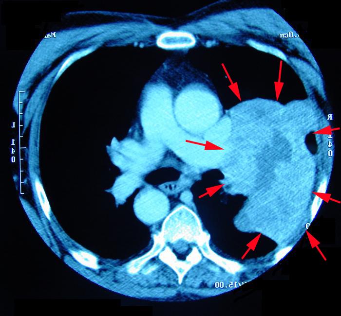
According to a study, from the University College London, Transvaginal ultrasound features high specificity and sensitivity for diagnosing endometrial cancer. The researchers discovered that transvaginal ultrasound is useful for high-risk groups prone to endometrial cancer and especially in the management of postmenopausal females subjecting to pelvic scans for causes different from vaginal bleeding.

The most common gynecological cancer in Europe is endometrial cancer, and in many northern and western European countries it has a rising occurrence in postmenopausal females. Overall occurrence of endometrial cancer in the U.K increased from 13.5 per 100,000 in 1993 to 17.9 per 100,000 in 2005.
In order to read the rest of the article please visit:
Transvaginal Ultrasound Is Useful For Detecting Endometrial Cancer, Study

Sunday 26 December 2010

Nordion Ships The First Mo-99 Sample From Russia.

Nordion Inc., a major company providing services and products to healthcare market across the world, announced that the first sample of its Molybdenum-99 (Mo-99) has been shipped from "Isotope", Nordion’s partner in Russia. Mo-99 and other Medical isotopes are used by a wide range of hospitals and healthcare facilities in order to diagnose and treat several medical conditions, such as cardiac and neurological disorders in addition to several forms of cancer.
Back in September, Nordion and Isotope made a supply agreement for Mo-99 that extends until 2020. The recently-shipped sample will be evaluated at Nordion’s facility in Ottawa, Canada. The assessment is taking place in order to get product registration and regulatory approvals before allowing Nordion to start supplying the medical isotope in the Canadian markets.

To read of this news, please visit:

Nordion Ships The First Mo-99 Sample From Russia.

New York Times Reported Uncomfortable Questions On Breast Cancer Screening Guidelines.

Mammography newsAccording to New York Times, debates have been taking place since the release of breast cancer screening recommendations over than one year ago; it was reported that there are still "lingering, uncomfortable questions about when and how often to undergo breast cancer screenings, and how to balance the benefits of early diagnosis with the harms of mammography”.

New guidelines were released by the U.S Preventive Services Task Force in November 2009, mentioning that females with an average risk of breast cancer should start imaging for the disease at age 50 other than females at age 40. The panel recommendations mentioned that the decision to begin imaging every two years "should be an individual one" that "takes patient context into account, including the patient's values regarding the specific benefits and harms."
In order to read the rest of the article please visit:
New York Times Reported Uncomfortable Questions On Breast Cancer Screening Guidelines.

EMRs Do Not Always Mean Higher Quality Of Care, Study.

A new research, published this week in the American Journal of Managed Care, found that the adoption of electronic medical records (EMR) in U.S. hospitals has limited effect on the quality of care.


The researchers at the nonprofit Rand analyzed the quality of care at 2,021 hospitals between 2003 and 2007 across three conditions: pneumonia, heart attack, and heart failure. The number of hospitals using either basic or advanced EMRs grew from 24 % in 2003 to 38 % in 2006.

To read the rest of the article, please visit:
EMRs Do Not Always Mean Higher Quality Of Care, Study.

Thursday 23 December 2010

Texas Breast Specialists Practice Awarded The ACR’s Breast Imaging Centre of Excellence.

Mammography newsThe American College of Radiology (ACR) has awarded Texas Breast Specialists, a related practice of Texas Oncology-Amarillo Cancer Center, the status of a “Breast Imaging Center of Excellence.”
Texas Specialists is considered to have the most developed breast screening equipment and the best diagnostic services in Amarillo. The practice provides all the screening techniques that can detect breast cancer in an earlier stage when the treatment is more effective, among these techniques, the digital screening and diagnostic mammography, breast-specific gamma imaging, breast ultrasound, stereotactic and ultrasound-guided needle and vacuum probe breast biopsies, ductography, and second opinion consultations.

To read the rest of the article, please visit:
 Texas Breast Specialists Practice Awarded The ACR’s Breast Imaging Center of Excellence.

iSoft Signs An Agreement With CliniWorks For Worldwide Distribution Of AccelFind.

iSoft, a leading Australian company providing healthcare IT solutions, announced that it has signed a new agreement with CliniWorks, a US-based healthcare IT marketing company, in order to distribute iSoft’s AccelFind software to hospitals and healthcare facilities throughout the world.
AccelFind allows its users to extract medical knowledge from any form or type of data. The knowledge can be obtained from sources such as free text notes, discharge summaries or other forms of data such as those available in electronic medical records (EMR). The recently-signed agreement will include iSoft offering its AccelFind among its Life Sciences solution, and it will provided under the ‘Powered by CliniWorks’ banner.

To read the rest of this news, please visit:
iSoft Signs An Agreement With CliniWorks For Worldwide Distribution Of AccelFind.

Philips Cooperates With German Partners To Improve MPI Technology.

Royal Philips Electronics, a major company providing medical imaging systems, has recently announced the beginning of a new German public-private partnership, which aims to improve the developments currently taking place in whole-body Magnetic Particle Imaging (MPI) systems, along with the preclinical hybrid systems that use a combination of MPI and the traditional MRI scans.

The German Federal Ministry of Education and Research committed to offer a fund of EUR 10.6 million to the consortium partners, and the total budget will be EUR 20.3 million. Philips aims to strengthen its position as a leading provider of the latest medical imaging systems. Therefore, the company is contributing the researches carried out to develop MPI; the imaging technique was designed by scientists at Philips.
In order to read the rest of the article please visit:
Philips Cooperates With German Partners To Improve MPI Technology.

Wednesday 22 December 2010

New Breast MRI Imaging and Biopsy System Presented By WCA Hospital

WCA Hospital is pleased to provide Breast MRI Imaging and MRI-guided Breast Biopsy - a minimally invasive procedure in which a breast biopsy is guided by highly detailed magnetic resonance imaging. When Breast MRI scans are used along with conventional mammography, they offer valuable data for the classification and the diagnosis of breast disease.

Breast Cancer in the United States is considered the most common kind of cancer among females and also the second leading cause of death in females. But, as the result of the diagnosis in an early stage and the development in treatment, a lot of females today are cured from breast cancer. Betsy T. Wright, WCA Hospital President/CEO  mentioned "Our new technological capabilities extends the range of quality diagnostic services that WCA offers and broadens our commitment to providing our patients with access to the best possible care in this region,"
In order to read the rest of the article please visit:
New Breast MRI Imaging and Biopsy System Presented By WCA Hospital

Brain Scan Can Predict When Dyslexic Children Can Read, Study.

A new study mentioned that by a simple brain scan researchers can predict when children with dyslexia will learn to read. It is known that brain disorder makes it very hard to children even clever ones to learn how to read, which is considered as a critical reason for frustration.
Improvements of children’s reading skills can be predicted accurately by brain scans with an accuracy of 90%, according to a research published in the Proceedings of the National Academy of Science (PNAS).

To read the rest of the article,please visit:
Brain Scan Can Predict When Dyslexic Children Can Read, Study.

Tuesday 21 December 2010

TMI Launches Wireless Foot/Hand Switch For Mobile C-Arms And Cardiovascular Systems.

TransAmerican Medical Imaging (TMI) has announced the launch of the Spectre, a wireless foot switch and hand switch system for GE-OEC Series 9800 and 9900 mobile C-Arms, and the Philips Cardiovascular product line. 
Bob Woodward, TMI’s President, explaining the capabilities of Spectre: “The TMI Spectre replaces the hard-wired foot and hand switches found on GE-OEC Series 9800™ and 9900™ systems, and on Philips fixed Cardiovascular systems. The Spectre eliminates the most common problems found with wired controls: dirty and hard-to-clean cables on the floor, broken cables and it allows OR staff to operate a system from any convenient position near the X-Ray source. Since cables on the floor are also a tripping hazard, this new wireless technology is an effective safety improvement.”
The Spectre wireless system will operate within a radius of 50 feet from a connected system. Since it is not infra-red, it does not have to be “line of Sight”. It can also operate for 20 consecutive hours without the need to be charged. Recharging takes only 2-4 hours.

To read the rest of this news, please visit:

TMI Launches Wireless Foot/Hand Switch For Mobile C-Arms And Cardiovascular Systems.

Diopsys® CORDA™ New Imaging Software To Help Ophthalmologists

New developed software that is using existing Spectral Domain Optical Coherence Tomography (SD-OCT) images will be released soon by Diopsys, Inc. This developed imaging software is going to help ophthalmologists and optometrists in procedures carried out for diagnosing retinal and optic nerve diseases like glaucoma, through providing them with thickness measurements of the Retinal Nerve Fiber Layer (RNFL). Moreover, this thickness may be affected in improper way by retinal vessels beside other things within the RNFL
Ophthalmologists and optometrists can reach for more accurate reading of the RNFL health from their OCT images, by using the new software (Diopsys® CORDA™) as it can analyze the inner building of the RNFL by the discrimination of its several components. So they can detect the disease and the vision disorders easily, which will be better for the sake of the patient.

Foe more info. please visit:

Diopsys® CORDA™ New Imaging Software To Help Ophthalmologists

Radiation Risks Might Be Overestimated, Study.

Despite the dominant trend indicating that radiation exposure during diagnostic imaging carries various risks for developing cancer, a recent report presented December 2 at RSNA 2010 demonstrated that risks may be lower than expected.
Researchers at Stanford University conducted a retrospective study of CT exposure using Medicare claims from 1998 through 2005. The research calculated the ionizing radiation exposure of the exams and estimated the associated cancer risks.
To read the rest of the article, please visit:
Radiation Risks Might Be Overestimated, Study.

Two-photon Microscopy Technique To Monitor Immune Cells In Lung, Study.

According to UCSF-led study in mice, researchers improved a way to equalize living lung tissue for screening without disrupting the normal function of the organ. This way was used for the first time and it permitted the researching team to observe both the unfolding events in the immune response during lung injury and the live interaction of living cells in the context of their environment. The authors mentioned that, the capability to screen the lung and other organs with minimum tissue disruption permits scientists to look deeper into many physiological aspects of injury and diseases for instance, cancer and diabetes. 

Senior author Max Krummel, PhD, UCSF associate professor of Pathology, whose lab improved the recent screening technique for viewing minute details of cellular interaction in tissues, reported "The nature of disease is complex, so if scientists can observe in real-time what's happening in tumors or immune responses as they occur, we can find new ways to intervene. We figured out a method for holding cells still enough to image them without interrupting their normal processes. This enabled us to observe cellular events as they happen naturally rather than the usual way, which is to stop the motion of cellular processes in order to photograph them."
In order to read the rest of the article please visit:

Two-photon Microscopy Technique To Monitor Immune Cells In Lung, Study.

Monday 20 December 2010

Cone Beam CT To Aid Mammography, Study.

At the Radiological community of North America Annual Scientific Assembly in Chicago, Avice O'Connell, M.D, URMC radiologist, discussed the studies concerning the Cone Beam Computed Tomography (CBCT) scanner in order to simplify it.
The CBCT offers high-quality pictures, besides, it is very important as it is easier on females because it permits breast scans in a significant way regardless of the need for pulling, compressing and flattening the breast tissue. O’ Connell is interested about its progress.
Moreover, a primary research noted that the breast coverage and radiation dose were in a comparison with mammography. However, the researchers didn’t imagine that the CBCT couldn’t replace mammography like a frontline screening tool. Besides, a lot of studies were necessary in order to illustrate its best usage in the clinic.

To read the rest of this news, please visit:

Cone Beam CT To Aid Mammography, Study.

DICOM Index Tracker Designed At Mayo Clinic To Track Radiation Doses.

Mayo Clinic, in Rochester, Minnesota, announced recently that is about to establish a new imaging exam tracking system, the DICOM Index Tracker (DIT). This system will allow a comprehensive view of the radiation doses delivered to patients over time. DTI is among the first of its type, as it replaces manual tracking system with a new computerized one, reducing the time needed for collecting radiation dose delivery information at various medical facilities.

The DICOM Index Tracker was created by Steve G. Langer, Ph.D. at Mayo Rochester; it is currently under further development at Mayo Clinic in Arizona in collaboration with Arizona State University. DIT is able to track all the information based on the imaging procedures applied on a patient, such as radiation dose, scanner utilization, and several other types of information. All this information are gathered and prepared for access by authorized personnel later on.
In order to read the rest of the article please visit:
DICOM Index Tracker Designed At Mayo Clinic To Track Radiation Doses.

Online Courses For Nurses By GE Healthcare

GE Healthcare has lately released a new precious library; open for subscription, of online courses and materials for nurses.The courses are directed mainly to general practice registered nurses and also to nurses specialized in other fields involving, telemetry, obstetrics beside emergency and critical care.
To read the rest of the article please visit:
Online Courses For Nurses By GE Healthcare

Sunday 19 December 2010

The Risk Of Thyroid Cancer Increased After Subjection Of Children To Radiation.

According to the University of Rochester Medical Center research, when children are subjected to head and neck radiation, whether as a result of multiple diagnostic CT scans or cancer treatment, it causes the increase of risk of thyroid cancer along the next fifty-eight years or more.

The study was published in the December 2010 edition of the journal, Radiation Research, it involved a group of children subjected to medical radiation and followed up for thyroid cancer incidence. The data obtained through the study might offer some information related to the rates of thyroid cancer, why the rates continue to increase as the general public through more used screening exams for instance computed tomography (CT) is increasingly subjected to higher doses of radiation.
In order to read the rest of the article please visit:
The Risk Of Thyroid Cancer Increased After Subjection Of Children To Radiation

Prostate Cancer With A Single Dose Radiation, Study.

Radiation oncologists at Beaumont Hospital in Michigan have conducted a study to evaluate a new treatment for men suffering from prostate cancer with a single dose of radiation to diminish the risk of it to a lower level.


The first single dose treatment in the world has been performed by Alvaro Martinez, M.D., FACR, professor and chair, Oakland University William Beaumont School of Medicine, department of Radiation Oncology, Beaumont Hospitals.The study is based on 18 years of researches by Dr. Martinez and other Beaumont radiation oncologists aiming at destroying cancer cells with very short-term radiation treatment.
To read the rest of the article please visit:
Prostate Cancer With A Single Dose Radiation, Study.

Thursday 16 December 2010

PET Scans Used For Better Understanding Of Alzheimer's Disease

Researchers from The Karolinska Institute, Sweden, revealed in the medical journal Brain that it was found, for the first time, through an Alzheimer's disease  patient who was detected to have amyloids while conducting PET scans on his brain regularly as his disease progressed, and then his brain was analyzed after he died. The researchers said that the study gave important data on the pathological course of Alzheimer's disease. 
Alzheimer's disease, which also known as SDAT (Senile Dementia of the Alzheimer Type), is a serious neurologic brain disease that results in irreversible loss of neurons and intellectual abilities such as reasoning and memory. Brain cells die due to the developed plaques in the structure of the brain during the course of the disease. "The method of forming the plaques in the brain and whether they are the main reason of Alzheimer's are still unknown" the authors explained.

To read the rest of this news, please visit:

PET Scans Used For Better Understanding Of Alzheimer's Disease

Advanced Radiology Presents A New Open 1.2T MRI

Recently, Advanced Radiology, the largest medical imaging provider in the state of Maryland, has presented Baltimore area's first 1.2 Tesla High Field Open MRI, thanks to the new system, patients and physicians will not have to sacrifice image quality when using an Open MRI. Patients who cannot be subjected to MRI in a traditional closed-bore unit as a result of claustrophobia or height/weight limitations, can now use the new high-strength, boreless magnet as it is considered a major improvement for those patients.
While having an acceptable picture quality in comparison with 1.5 Tesla MRIs, Claustrophobic patients can avoid receiving sedatives or anesthesia. Advanced Radiology's new magnet has created more comfortable and convenient experience for patients, while offering highly precise images that can help in detecting orthopedic, body and other disorders in an accurate way.
in order to read the rest of the article please visit:
http://www.healthimaginghub.com/126-medical-imaging/2521-advanced-radiology-presents-a-new-open-12t-mri.html

SPECT-CT Can Reduce Lymphedema In Breast Cancer Patients.

According to the radiation oncologists at Mayo Clinic, they were capable of decreasing the number of lymph nodes critical for removing fluid from the arm that subjected to damaging radiation doses by 55% with the utilization of a specialized scan.


The researchers reported that integrating single photon emission computed tomography (SPECT) with the computerized tomography (CT) scans used for breast cancer radiotherapy planning may provide patients substantial protection against lymphedema, chronic swelling of tissue and an incurable that results from damage to lymph nodes sustained during breast cancer radiation.

To read the rest of the article, please visit:
SPECT-CT Can Reduce Lymphedema In Breast Cancer Patients.

Wednesday 15 December 2010

Ultrasound Not Needed For Medical Abortion, Study.

Despite a lot of females looking for medical abortion usually undergo an ultrasound scan, a new study is indicating that this procedure may not necessary for them. The findings of the study were published in the British Journal of Obstetrics and Gynecology. Medical abortion is usually carried out by giving the pregnant female a dose of mifepristone and then another one of misoprostol. This type of abortion in conducted in the United States within nine weeks of a female's last menstruation.

To read the rest of this news, please visit:

Ultrasound Not Needed For Medical Abortion, Study.

WellDoc Integrates mHealth Diabetes Coaching Platform Into An EHR System.

WellDoc, a leading healthcare company innovating technology solutions to improve chronic disease management outcomes and reduce healthcare costs, has revealed today the first integration of a clinically tested mHealth coaching platform with a leading electronic health records (EHR) system.

The integration of the WellDoc DiabetesManager solution into an EHR will ease and enhance patient care by combining a best-in-class patient coaching platform and clinical decision support with the workflow efficiency of an EHR system.

For more info. please visit:


WellDoc Integrates mHealth Diabetes Coaching Platform Into An EHR System.

Ultrasound Is Used To Detect Endometrial Cancers.

Researchers found that transvaginal ultrasound screening can show many cases of endometrial cancer in postmenopausal women before any appearance of symptoms. According to Ian Jacobs, MD, of University College London, and colleagues more than 80% of cases can be determined by this test and about 15% of them is false. 
Jacobs and colleagues reported online in The Lancet Oncology that in order to develop the sensitivity and specificity, the ultrasound screening would be restricted to a high-risk population. Many experts disagree with this technique as it can't be cost-effective.  Jonathan Berek, MD, of Stanford University School of Medicine in Stanford, Calif, said in an e-mail "Symptoms usually vaginal bleeding will be improved in all women with endometrial cancer before it spreads. Once symptoms appear the diagnosis will be performed by an endometrial biopsy".
In order to read the rest of the article please visit:
Ultrasound Is Used To Detect Endometrial Cancers.

MRI Used For Imaging Birth Process In Germany.

Women's imaging news Magnetic Resonance Imaging (MRI) is one of the most important diagnostic imaging techniques, as it offers highly detailed images in a very safe procedure, as the imaging technique does not involve radiation. Recently, MRI scans were used to generate images of a baby while being born. The imaging procedure took place at Charite Hospital in Berlin, Germany.

To read the rest of the article, please visit:
MRI Used For Imaging Birth Process In Germany.

Sunday 12 December 2010

Researchers Use Patient's Own Blood to Treat Hamstring Injury

CHICAGO—Researchers in London say they have found an effective two-part treatment for microtears in the hamstring: injections of the patient's own blood and a steroid along with "dry-needling," in which repeated needle punctures cause controlled internal bleeding in the injured area. Results of the study were presented today at the annual meeting of the Radiological Society of North America (RSNA).
RSNA Hamstring Enthesopathy
A magnetic resonance (MR) image depicting increased signal on a fat saturated sequence to confirm hamstring complex enthesopathy—tendinopathy. Image and caption courtesy of the RSNA.
"By injecting the patient's own blood where it is needed at the site of a damaged tendon, we help the patient heal themselves," said lead researcher Waseem A. Bashir, M.D., a radiologist at Royal National Orthopaedic Hospital and Ealing Hospital in London. "Blood contains many growth factors, and the injections have been shown to promote faster healing of certain injuries."

To read the rest of the article please visit:
Researchers Use Patient's Own Blood to Treat Hamstring Injury

Thursday 9 December 2010

CT Best at Uncovering Drug Mule Payload

CHICAGO—According to a study presented today at the annual meeting of the Radiological Society of North America (RSNA), the best way to detect cocaine in the body of a human drug courier, known as a mule, is through the use of CT.


"Cocaine from South America is making its way to Europe through Africa," said Patricia Flach, M.D., a radiologist at University Hospital of Berne and Institute of Forensic Medicine of Berne in Switzerland. "From Africa, drug mules most commonly try to enter the European Union and Switzerland.
In order to read the rest of the article please visit:
CT Best at Uncovering Drug Mule Payload

People With Sleep Apnea at Higher Risk for Aggressive Heart Disease

CHICAGO—People with obstructive sleep apnea (OSA), a sleep disorder associated with obesity, have more non-calcified or "bad" plaque in their coronary arteries, according to a study presented today at the annual meeting of the Radiological Society of North America (RSNA).
RSNA Sleep Apnea
A CT image of individual heart vessels showing calcified atherosclerotic plaque (arrows) and abundant non-calcified ("soft") plaque (arrowheads) in a patient with obstructive sleep apnea. Image and caption courtesy of the RSNA.
"Our study reveals that individuals with obstructive sleep apnea are prone to developing an aggressive form of atherosclerosis that puts them at risk for impaired blood flow and cardiovascular events," said U. Joseph Schoepf, M.D., professor of radiology and medicine and director of cardiovascular imaging at the Medical University of South Carolina in Charleston, S.C.

To raed the rest of the article please visit:
People With Sleep Apnea at Higher Risk for Aggressive Heart Disease

Wednesday 8 December 2010

Expert Panel Addresses Safety in Medical Imaging

CHICAGO—An expert panel convened today at the annual meeting of the Radiological Society of North America (RSNA) to discuss medical imaging appropriateness, ionizing radiation from imaging procedures, and efforts under way to curb overutilization, decrease radiation dose, and educate patients on the risks and benefits of medical imaging.

Panel members included:
    RSNA2010 Imaging Safety James A. Brink
    James A. Brink, M.D. Image courtesy of Yale University School of Medicine.
  • James A. Brink, M.D., professor and chairman of the department of diagnostic radiology at Yale University School of Medicine in New Haven, Conn., and co-chairman of the joint Adult Radiation Protection Task Force cosponsored by the RSNA and the American College of Radiology (ACR).
  • William R. Hendee, Ph.D., distinguished professor of radiology, radiation oncology, biophysics and bioethics at the Medical College of Wisconsin in Milwaukee.
  • Christoph Wald, M.D., Ph.D., executive vice-chairman of the department of radiology at Lahey Clinic in Burlington, Mass., and associate professor of radiology at Tufts University Medical School in Boston; and
  • Moderator Mary C. Mahoney, M.D., professor of radiology and director of Breast Imaging at the University of Cincinnati Medical Center and chair of the RSNA Public Information Committee.

To read the rest of this news, please visit:


Expert Panel Addresses Safety in Medical Imaging

New Study Reports Effects of Endurance Running

CHICAGO—Using a mobile MRI unit, researchers followed runners for two months along a 4,500-kilometer course to study how their bodies responded to the high-stress conditions of an ultra-long-distance race, according to a study presented today at the annual meeting of the Radiological Society of North America (RSNA).
RSNA Endurance Running
A series of MR images of the brain showing loss of gray matter volume. Image and caption courtesy of the RSNA.
"Due to the exceptional setting of this study, we could acquire huge amounts of unique data regarding how endurance running affects the body's muscle and body fat," said Uwe Schütz, M.D., a specialist in orthopedics and trauma surgery in the department of diagnostic and interventional radiology at the University Hospital of Ulm in Germany. "Much of what we have learned so far can also be applied to the average runner."

To read the rest of the article, please visit:

New Study Reports Effects of Endurance Running

Annual Breast Cancer Screening Beginning at Age 40 Reduces Mastectomy Risk

CHICAGO—Having a yearly mammogram greatly reduces the risk of mastectomy following breast cancer in women between the ages of 40 and 50, according to a study being presented today at the annual meeting of the Radiological Society of North America (RSNA).


"The results of this study support the importance of regular screening in the 40 to 50 age group," said lead author Nicholas M. Perry, M.B.B.S., F.R.C.S., F.R.C.R., director of The London Breast Institute at The Princess Grace Hospital in London. "Women in this age group who had undergone mammography the previous year had a mastectomy rate of less than half that of the others."
In order to read the rest of the article please visit:
http://www.healthimaginghub.com/rsna-2010/2428-annual-breast-cancer-screening-beginning-at-age-40-reduces-mastectomy-risk.html

Belly Fat Puts Women at Risk for Osteoporosis

CHICAGO—For years, it was believed that obese women were at lower risk for developing osteoporosis, and that excess body fat actually protected against bone loss. However, a study presented today at the annual meeting of the Radiological Society of North America (RSNA) found that having too much internal abdominal fat may, in fact, have a damaging effect on bone health.
"We know that obesity is a major public health problem," said the study's lead author, Miriam A. Bredella, M.D., a radiologist at Massachusetts General Hospital and assistant professor of radiology at Harvard Medical School in Boston. "Now we know that abdominal obesity needs to be included as a risk factor for osteoporosis and bone loss."

For more details please visit:
Belly Fat Puts Women at Risk for Osteoporosis

Monday 6 December 2010

Diagnosis Uncertainty Increases Anxiety in Patients…

CHICAGO—Have you ever felt uneasy sitting in a doctor's waiting room or climbed the walls waiting for your test results? That feeling of anxious uncertainty can be more stressful than knowing you have a serious illness, according to a study presented today at the annual meeting of the Radiological Society of North America (RSNA).

"Not knowing your diagnosis is a very serious stressor," said the study's lead author, Elvira V. Lang, M.D., associate professor of radiology at Harvard Medical School in Boston. "It can be as serious as knowing that you have malignant disease or need to undergo a possibly risky treatment."

To read the rest of the article, please visit:

Diagnosis Uncertainty Increases Anxiety in Patients…

Burton P. Drayer, M.D., Named RSNA President

CHICAGO—Burton P. Drayer, M.D., was named president of the Radiological Society of North America (RSNA) Board of Directors today at the Society's annual meeting.
Dr. Drayer is executive vice president for Risk at The Mount Sinai Medical Center in New York City, and since 1995, has served as the Dr. Charles M. and Marilyn Newman Professor and chairman of the department of radiology at The Mount Sinai School of Medicine. He served as president of The Mount Sinai Hospital from November 2003 to September 2008.
As president, Dr. Drayer is committed to RSNA's role in supporting education, research, information technology and patient safety.
"My goals for 2011 are to enhance collaborations among radiology and non-radiology societies, as well as to extend the utilization and recognition of the RSNA's extensive technology platform," he said. "I hope to accelerate our critically important research agenda by fostering the growth and quality of our journals, annual meeting program, and Research & Education (R&E) Foundation grant support, and I am committed to expanding the role that RSNA plays in advancing radiation safety."
Dr. Drayer received his undergraduate degree in political science from the University of Pennsylvania in Philadelphia. In 1971, he received his medical degree from Chicago Medical School and went on to complete a medical internship and neurology residency at the University of Vermont in Burlington. Dr. Drayer served his radiology residency followed by a neuroradiology fellowship at the University of Pittsburgh Medical Center.

To read the rest of this news, please visit:

Burton P. Drayer, M.D., Named RSNA President

Virtual Biopsy May Allow Earlier Diagnosis of Brain Disorder in Athletes

CHICAGO—In a study of retired professional athletes, researchers found that a specialized imaging technique called magnetic resonance spectroscopy (MRS) may help diagnose chronic traumatic encephalopathy (CTE), a disorder caused by repetitive head trauma that currently can only be definitively diagnosed at autopsy. Results of the study were presented today at the annual meeting of the Radiological Society of North America (RSNA).


"The devastating effects of brain injuries suffered by professional football players who repeatedly suffered concussions and sub-concussive brain trauma during their careers have put the spotlight on CTE," said Alexander P. Lin, Ph.D., a principal investigator at the Center for Clinical Spectroscopy at Brigham and Women's Hospital in Boston. "However, blows to the head suffered by all athletes involved in contact sports are of increasing concern."
In order to read the rest of the article please visit:
Virtual Biopsy May Allow Earlier Diagnosis of Brain Disorder in Athletes

Acupuncture Changes Brain's Perception and Processing of Pain

CHICAGO—Using functional magnetic resonance imaging (fMRI), researchers have captured pictures of the brain while patients experienced a pain stimulus—with and without acupuncture—to determine acupuncture's effect on how the brain processes pain. Results of the study, which the researchers say suggest the effectiveness of acupuncture, were presented today at the annual meeting of the Radiological Society of North America (RSNA).
"Until now, the role of acupuncture in the perception and processing of pain has been controversial," said lead researcher Nina Theysohn, M.D., from the department of diagnostic and interventional radiology and neuroradiology at University Hospital in Essen, Germany. "Functional MRI gives us the opportunity to directly observe areas of the brain that are activated during pain perception and see the variances that occur with acupuncture." fMRI measures tiny metabolic changes that take place in an active part of the brain, while a patient performs a task or is exposed to a specific external stimulus.

To read the rest of the article please visit:
Acupuncture Changes Brain's Perception and Processing of Pain

Sunday 5 December 2010

RSNA Presents Special Presidential Award to Hans G. Ringertz, M.D., Ph.D.

CHICAGO—Today the Radiological Society of North America (RSNA) presented its Special Presidential Award to Hans G. Ringertz, M.D., Ph.D., of Linköping, Sweden. The RSNA reserves the Special Presidential Award for individuals who have made significant contributions to the field of radiology or the radiologic sciences.


During his nearly 50 years in medicine, Dr. Ringertz has worked tirelessly to preserve, defend, expand, and advance radiology.

"Professor Ringertz is the proverbial man for all seasons," said 2010 RSNA President Hedvig Hricak, M.D., Ph.D., Dr. h.c. "He is the pioneer of pediatric MR imaging, a world leader in radiation safety, and he headed one of the most prestigious university radiologic departments in the world. His membership and presidency of the Nobel committee always ensured biomedical imaging a fair review."

Since 2006, Dr. Ringertz has been a professor of radiology at Linköping University Hospital in Linköping, Sweden, and chairman of the board for the university's Centre for Medical Imaging Science and Visualization. He was a professor and chairman of radiology at the Karolinska Institute in Stockholm from 1984 to 2006 and has served as a professor emeritus since.
In order to read the rest of the article please visit:
RSNA Presents Special Presidential Award to Hans G. Ringertz, M.D., Ph.D.

Sarah S. Donaldson, M.D., Named RSNA Chairman of the Board

CHICAGO—Sarah S. Donaldson, M.D., was named chairman of the Radiological Society of North America (RSNA) Board of Directors today at the Society's annual meeting.
Dr. Donaldson has served as associate chair of the Department of Radiation Oncology at Stanford University School of Medicine in Stanford, Calif., since 1997, where she is also the Catherine and Howard Avery Professor of Radiation Oncology. In addition, she serves as deputy clinic chief and associate residency program director of radiation oncology at Stanford Hospital and Clinics. She is chief of radiation oncology service and a member of the medical board at Lucile Salter Packard Children's Hospital at Stanford, as well as a member of the medical staff of the VA Palo Alto Health Care System.
"My primary goal as chairman of the RSNA board will be to initiate a comprehensive review of the RSNA strategic plan," Dr. Donaldson said. "I will focus on education via the Internet, the international needs of radiologists, quality improvements in our field, and strengthening RSNA's interactions with sub-specialty societies. My intent is to listen and to hear the voices of all radiologists and address their concerns, while focusing on the issues identified as central to the RSNA strategic plan."

To read the rest of the news please visit:
Sarah S. Donaldson, M.D., Named RSNA Chairman of the Board

Light Exercise May Prevent Osteoarthritis

CHICAGO — People at risk for osteoarthritis may be able to delay the onset of the disease or even prevent it with simple changes to their physical activity, according to a study presented today at the annual meeting of the Radiological Society of North America (RSNA).
RSNA Osteoarthritis

"According to the results of our study, participating in a high-impact activity, such as running, more than one hour per day at least three times a week appears associated with more degenerated cartilage and potentially a higher risk for development of osteoarthritis," said the study's senior author Thomas M. Link, M.D., professor of radiology and chief of musculoskeletal imaging at the University of California, San Francisco (UCSF). "On the other hand, engaging in light exercise and refraining from frequent knee-bending activities may protect against the onset of the disease."
To view more info. please visit:
http://www.healthimaginghub.com/rsna-2010/2405-light-exercise-may-prevent-osteoarthritis.html

Thursday 2 December 2010

Image Wisely Initiative to Help Providers Reduce Radiation Dose

As medical imaging exams have replaced more invasive procedures, benefiting patients and revolutionizing medicine, people's exposure to medical radiation has increased. Imaging providers are concerned that radiation exposure over time could have adverse effects for patients. They continue to act to ensure that patients receive only necessary scans and to reduce radiation dose used in these exams.

For more info. , please visit:
http://www.healthimaginghub.com/rsna-2010/2404-image-wisely-initiative-to-help-providers-reduce-radiation-dose.html

George S. Bisset III, M.D., Named RSNA President-Elect

CHICAGO - George S. Bisset III, M.D., was named president-elect of the Radiological Society of North America (RSNA) Board of Directors today at the Society's annual meeting. Dr. Bisset is currently chief of pediatric radiology at Texas Children's Hospital, as well as professor of radiology and Edward B. Singleton Chair of Pediatric Radiology at Baylor College of Medicine in Houston.
"I am looking forward to my tenure as president-elect, during which time I will also serve as secretary-treasurer," Dr. Bisset said. "The financial management of the RSNA is in excellent hands. I can only aspire to adding some new perspectives on improving our portfolio and providing some new insight into means for building our membership and membership programs."
Dr. Bisset earned his medical degree in 1975 at the University of South Florida College of Medicine. At Cincinnati Children's Hospital Medical Center (CCHMC), Dr. Bisset completed his pediatric residency in 1977 and a fellowship in pediatric cardiology in 1980. In 1983, he began his radiology residency at the University of Cincinnati Medical Center, combined with a pediatric radiology fellowship at CCHMC, completing both in 1986.

To read the rest of this news, please visit:

http://www.healthimaginghub.com/rsna-2010/2438-george-s-bisset-iii-md-named-rsna-president-elect.html

Screening Tool May Better Identify Heart Disease in African Americans

CHICAGO—In a study presented today at the annual meeting of the Radiological Society of North America (RSNA), researchers say they may have an explanation as to why African Americans, despite having lower amounts of coronary artery calcification, are at increased risk for heart attacks and other cardiovascular events compared with Caucasians.
The answer, according to researchers at the Medical University of South Carolina (MUSC) in Charleston, S.C., may be increased levels of non-calcified plaque, which consists of buildups of soft deposits deep in the walls of the arteries that are not detected by some cardiac tests. Non-calcified plaque is more vulnerable to rupturing and causing a blood clot, which could lead to a heart attack or other cardiovascular event.


"The African Americans and Caucasians we studied had approximately the same amount of plaque in their arteries, but different kinds of plaque," said John W. Nance Jr., M.D., a researcher in the department of radiology and radiological sciences at MUSC.
For more information please visit: http://www.healthimaginghub.com/rsna-2010/2413-screening-tool-may-better-identify-heart-disease-in-african-americans.html

Cancer Risk From Medical Radiation May Have Been Overestimated

CHICAGO—The risk of developing radiation-induced cancer from CT may be lower than previously thought, according to a study presented today at the annual meeting of the Radiological Society of North America (RSNA).

"Radiation from medical imaging has gotten a tremendous amount of attention in recent years," said Aabed Meer, an M.D. candidate at Stanford University in Palo Alto, Calif. "This is one of the first studies to track CT utilization in such a large population."

The researchers conducted a retrospective study using Medicare claims from 1998 through 2005 to analyze the distribution of CT scans, determine the ionizing radiation exposure associated with the exams and estimate the associated cancer risk in a population of older adults.

For more information please visit : http://www.healthimaginghub.com/rsna-2010/2425-cancer-risk-from-medical-radiation-may-have-been-overestimated.html.

Diagnosis Uncertainty Increases Anxiety in Patients

CHICAGO—Have you ever felt uneasy sitting in a doctor's waiting room or climbed the walls waiting for your test results? That feeling of anxious uncertainty can be more stressful than knowing you have a serious illness, according to a study presented today at the annual meeting of the Radiological Society of North America (RSNA).
"Not knowing your diagnosis is a very serious stressor," said the study's lead author, Elvira V. Lang, M.D., associate professor of radiology at Harvard Medical School in Boston. "It can be as serious as knowing that you have malignant disease or need to undergo a possibly risky treatment."
To read the rest of this article, please visit our website:
http://www.healthimaginghub.com/rsna-2010/2401-diagnosis-uncertainty-increases-anxiety-in-patients.html