 very low when using mammography as the radiation dose in this procedure is small. The study is highlighted in October issue of Radiology. R. Edward Hendrick, clinical professor of radiology at the University of Colorado-Denver, School of Medicine in Aurora, Co., the lead author of the study, said "A single breast-specific gamma imaging (BSGI) or positron emission mammography (PEM) examination carries a lifetime risk of inducing fatal cancer greater than or comparable to a lifetime of annual screening mammography starting at age 40,"
Thursday, 26 August 2010
BSGI And PEM Have Higher Risks For Radiation-induced Cancer
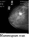
According to a new study, nuclear-based breast imaging procedures, such as BSGI and PEM, are capable of elevating the risk of radiation-induced cancer. Yet, such risk is very low when using mammography as the radiation dose in this procedure is small. The study is highlighted in October issue of Radiology. R. Edward Hendrick, clinical professor of radiology at the University of Colorado-Denver, School of Medicine in Aurora, Co., the lead author of the study, said "A single breast-specific gamma imaging (BSGI) or positron emission mammography (PEM) examination carries a lifetime risk of inducing fatal cancer greater than or comparable to a lifetime of annual screening mammography starting at age 40,"
very low when using mammography as the radiation dose in this procedure is small. The study is highlighted in October issue of Radiology. R. Edward Hendrick, clinical professor of radiology at the University of Colorado-Denver, School of Medicine in Aurora, Co., the lead author of the study, said "A single breast-specific gamma imaging (BSGI) or positron emission mammography (PEM) examination carries a lifetime risk of inducing fatal cancer greater than or comparable to a lifetime of annual screening mammography starting at age 40,"
New Mobile Mammography Unit In West Sacramento
Breast cancer is one of the most common forms of cancer between women. However, the disease has high care rate and overall all survival rate provided that the condition is detected early. Realizing this, West-St. Joseph's Medical Center and the Safeway Foundation have combined their efforts to offers women in West Sacramento and Rancho Cordova a new mobile mammography screening service, which will start next week.
TeraMedica Launches - Enterprise Data Migration Services (EDM), migrating DICOM and beyond
TeraMedica, a Milwaukee-based, medical informatics company, announced the launch of its new Enterprise Data Migration (EDM) Services offering as part of its commitment to solving the growing data migration and interoperability challenges across the enterprise according to CEO & President, Jim Prekop.
3D MRI Offers New Options In Pediatric Imaging At Stony Brook Medical Center
MRI has been one of the significantly important medical imaging techniques. The procedure offers highly-detailed images with high safety profile as it does not involve radiation  exposure. More advances and new applications for MRI are appearing every day. For instance, Jeffrey C. Hellinger, M.D., a pediatric imaging specialist at Stony Brook University Medical Center, mentioned that 3D MRI is a new medical imaging technique that can be used for visualizing fetal anatomy and detecting disorders in the womb in various clinical conditions. 3D MRI is highlighted in July-August issue of Applied Radiology.
exposure. More advances and new applications for MRI are appearing every day. For instance, Jeffrey C. Hellinger, M.D., a pediatric imaging specialist at Stony Brook University Medical Center, mentioned that 3D MRI is a new medical imaging technique that can be used for visualizing fetal anatomy and detecting disorders in the womb in various clinical conditions. 3D MRI is highlighted in July-August issue of Applied Radiology.
Subscribe to:
Comments (Atom)