 Agfa HealthCare, a major company providing medical imaging systems and healthcare IT solutions, has recently presented its latest innovation, IMPAX Kiosk, which represents a workflow improvement solution. IMPAX Kiosk is an interactive platform that permits the registration and check-in of patients to medical services at hospitals with no need for going to administration desks. Sarah Muckler, Senior Marketing Manager for Agfa HealthCare US, explained "For the patients, traditional check-in means waiting in a line before being afforded the opportunity to be personally checked-in by a receptionist. This can take fifteen minutes or more. With IMPAX Kiosk, we can reduce the check-in process time to five minutes." The use of the new systems will surely improve patients' satisfaction and will offer more time for administrative staff to concentrate on their other responsibilities.
Monday, 25 October 2010
Agfa Presents Its Latest Innovation, IMPAX Kiosk.
Agfa HealthCare, a major company providing medical imaging systems and healthcare IT solutions, has recently presented its latest innovation, IMPAX Kiosk, which represents a workflow improvement solution. IMPAX Kiosk is an interactive platform that permits the registration and check-in of patients to medical services at hospitals with no need for going to administration desks. Sarah Muckler, Senior Marketing Manager for Agfa HealthCare US, explained "For the patients, traditional check-in means waiting in a line before being afforded the opportunity to be personally checked-in by a receptionist. This can take fifteen minutes or more. With IMPAX Kiosk, we can reduce the check-in process time to five minutes." The use of the new systems will surely improve patients' satisfaction and will offer more time for administrative staff to concentrate on their other responsibilities.
CT, CTA, And Biopsy For Detection Of Natural Death Cause, Study
 According to a new study, researchers mentioned that using a combination of computed tomography (CT), postmortem CT angiography (CTA), along with biopsy can be useful as a minimally invasive procedure for detecting the natural causes of mortality, such as cardiac arrest. The study took place at the Institute of Forensic Medicine, Center for Forensic Imaging and Virtopsy, at the University of Bern in Bern, Switzerland. The findings of the study appear in the November issue of the American Journal of Roentgenology.
Africa Telehealth 2010 – Opening Sessions

Telehealth and telemedicine is currently one of the significantly growing fields in healthcare industry. This is due to the fact that both fields have various advantages that can truly improve the levels of healthcare and medical services. Africa Telehealth 2010 conference, currently taking place in Cairo, Egypt, is aiming to discuss the adoption of tele-health, the benefits and the challenges affecting that adoption. The event was organized by Texas Telehealth Tech (TTT) and sponsored by leading telemedicine solutions provider, Polycom & FVC, Gross Remote Conferencing, and emerging Telemedicine company in the Middle East, Telemed Providers. The event extends from 23th – 25th of October with Health Imaging Hub contributing as the official media sponsor. In this report, we highlight the events and sessions that took place during the first day of the conference.
The first day of Africa Telehealth 2010 included a number of highly interesting sessions. After welcoming the attendees, the event started with a presentation from Dr. Sahar Saleem, Radiology Professor, Faculty of Medicine (Kasr El Aini), Cairo University. She spoke about telemedicine in Egypt, mentioning that the first radiology practice in the country, also the first in Middle East, took place in 1922. Dr. Saleem briefly spoke about radiology statistics in Egypt, such as number of MRI units in the country (160), and their ratio in relation to population (2 MRI units per million capita). She also added that PACS systems are currently used in Egypt but on a very limited scale. Dr. Saleem confirmed during her presentation that telemedicine represents a suitable solution against a number of obstacles hindering the improvement of healthcare services, such as lack of specialists and equipment in rural areas, over population and over-crowded cities such as Cairo and Alexandria, where patients need a lot of time to reach radiology centers. Dr. Saleem added that telemedicine can improve the level of radiologists in Egypt as a result to continuous discussions and consultations with their colleagues in Western countries such as US and Canada, adding that such discussions can be useful if they take place between radiologists in Arab countries. She concluded that the expansion in telemedicine adoption in Egypt will be highly cost-effective, not only for patients, as they will no longer need to travel for long distances to radiology centers, but also for hospitals and healthcare facilities.
Monday, 18 October 2010
The Life - Saving Benefits Of Cardiac Imaging
Doctors depend on cardiac imaging when they have patients suffering from chest pain or other risk symptoms of heart problems. This imaging procedure helps doctors in finding out whether there is an evidence of heart disease, such as blockages in the coronary arteries or reduced blood flow to the heart.There are two methods of cardiac imaging including Cardiac Magnetic Resonance Imaging (MRI) and Cardiac Computed Tomography (CT) are allowing doctors to take a closer look at the heart and great vessels at little risk to the patient.
MRI uses huge magnets and radio-frequency waves to promote high-quality still and moving images of the body's internal structures; no X-ray exposure is included, Like anything MRI also has disadvantages : people with peacemakers cannot have MRIs. Also some people who are morbidly obese cannot fit in into an MRI system MRI scans require patients to hold still for extended periods of time. MRI exams can range in length from 20 minutes to 90 minutes or more. Even very slight movement of the part being scanned can cause distorted images which means the scanning will need to be repeated , while CT scan is an x-ray procedure which combines many x-ray images with the aid of a computer to generate cross-sectional views of the body. Cardiac CT uses advanced CT technology with or without intravenous iodine-based contrast to visualize cardiac anatomy, including the coronary arteries and great arteries and veins. CT scan has also disadvantages: Lack of IV access makes the CT procedure difficult to interpret, CTA is unsuitable when there is a large amount of existing coronary artery calcification.
To read the rest of the article, please visit The Life - Saving Benefits Of Cardiac Imaging
ACR Image Mertix Hosted An Event To Enhance Imaging Trials
 ACR image Mertix, an international imaging contract organization (CRO) including expertise in imaging trial design , techniques and data extraction, organized a recent event titled “The Novel Medical imaging Approaches for Early Drug Development Methods, technical issues and case studies” which took place at the at the Boston Marriott Cambridge, in Cambridge Massachusetts on the 16th of September.
Bruce Hillman, Chief scientific officer, M.D., FACR, of ACR Image Metrix, carried out a presentation titled, "Methodologic Considerations in Designing Pharmaceutical Trials Using Novel Imaging Methods."ACR Image Mertix is a leader in worldwide imaging trials. Industry experts from ACR Image Metrix offered great caliber research on ways and technical problems related to clinical trials. Mehdi Adineh , Ph.D., Scientific director of ACR Image Metrix imaging core laboratory, conducted a presentation named "Increasing the Effectiveness of Novel Imaging in Clinical Trials: The Role of the Imaging Core Laboratory,” while Greg Sorenson, a professor of radiology at Harvard Medical School, Massachusetts General Hospital ,M.D, had a presentation titled "Mechanistic Imaging in Cancer Trials: Lessons from Glioblastoma."
Monday, 11 October 2010
Hologic Offers 3D Mammography Training Program.
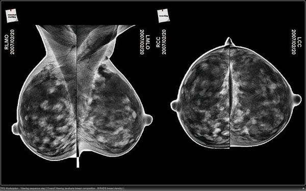 Any company should first design a program for physicians to exercise on how to read digital mammography files. Realizing this, Hologic, a major company providing breast imaging systems, decided to offer training on its new 3D digital mammography device before it becomes commercial product on the U.S. market.
NMR/MRI Used For Molecular Imaging Through Micro-fluidic Devices.
 In a recent study, Nuclear Magnetic Resonance (NMR) spectroscopy and Magnetic Resonance Imaging (MRI) were used to capture images of materials flowing through micro-fluidic "lab-on-a-chip" devices and focus on microscopic particles of special interest with new, unprecedented spatial and time resolutions. The study was conducted by chemist Alexander Pines and his researching team, in cooperation with the University of California (UC) at Berkeley and Lawrence Berkeley National Laboratory (Berkeley Lab). The study is highlighted in the journal Science, in a paper named "Zooming in on Microscopic Flow by Remotely Detected MRI."
Huntsville Memorial Hospital Awarded ACR’s Accreditation In Mammography
 As the result of a new survey, conducted by the American college of Radiology (ACR), Huntsville Memorial Hospital (HMH) has been awarded a three-year accreditation in mammography, the ACR, located in Reston, Va., grants accreditation to facilities that achieve high practice standards. The accreditation is awarded after a peer-review evaluation of the practice. Assessments are organized and conducted by wide-certified physicians and medical physicists who are expert in the scope.
Monongahela Valley Hospital Joins Image Radiology Group LP.
 In an announcement, Monongahela Valley Hospital declared that it has joined, Charleroi-based, Image Radiology Group LP. They are going to provide different diagnostic services at 2001 Lincoln Way, Oak Park Mall, White Oak and 4198 Washington Road, Route 19, McMurray. Louis J. Panza, Jr., president and CEO of Monongahela Valley Hospital mentioned that the partnership was effective stating from September 23 and services through the hospital could be offered as early as November. He added that "Image Radiology Group is well respected in the region and, like MVH, recognizes the importance of high-quality, prompt and professional service. We look forward to a great partnership."
A New Mammography Machine Added To McDuffie Regional Medical Center
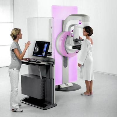 In March, McDuffie Regional Medical Center presented a new digital mammography system from Hologic. The new machine helps to detect breast cancer and to diagnose the disease earlier. Sheron Rutkowski, the director of radiology at MRMC, mentioned that "It's faster because we no longer have to develop film,” she added "The image just pops up within seconds on the computer screen. The patients no longer have to wait for us to develop the film before they can leave."
A Study Shows That FePt Nanoparticles Can Perform As CT/MRI
 In a recent study, published in the Sept. 29 issue of Journal of American Chemical Society, Iron-platinum alloy (FePt) nanoparticles can perform as dual modal CT/MRI molecular imaging contrast agent in clinical procedures. Chia-chun Chen, PhD, a professor in the department of chemistry at National Taiwan Normal University and his team has developed and used the water-solvable FePt nanoparticles of 3nm, 6 nm and 12 nm in diameter as dual modality contrast agent for CT/MRI molecular imaging.
Low-Intensity Pulsed Ultrasound Can Improve Poor Bone Healing.
 A new randomized controlled study published in the open access journal BMC Musculoskeletal Disorders, has showed that the use of low-intensity pulsed ultrasound (LIPUS) in patients suffering from tibial fractures with inadequate healing leaded to 34% greater bone mineral density (BMD) in the fracture area after 16 weeks than use of a sham device.
LIPUS to increase BMD.
Telemedicine Can Promote Early Diagnosis And Treatment Of Pediatric Heart Problems.
 New evidence that emphasizes the important role of telemedicine in promoting healthcare has been revealed by a recent study conducted by researchers from Children’s Mercy Hospitals and Clinics in Kansas City, Mo. The study showed that rapid electronic review of children's heart images by physicians approximately 200 miles away allowed earlier diagnosis and treatment of potentially serious pediatric cardiology problems. This result comes after an earlier report indicating that telemedicine could improve care for senior citizens battling depression.
Subscribe to:
Comments (Atom)