Newer MRI and CT-angiography proved nearly equal in identifying significant stenosis, and were equally accurate in predicting the need for revascularization in patients with known coronary artery disease (CAD), in the first direct comparison of MRA and CTA, say German researchers.
32-channel MRI offers equal accuracy in diagnosing stenosis with CTA, and is less invasive than conventional angiography.
In their study, the researchers led by Dr. Ashraf Hamdan, of the German Heart Institute, compared the diagnostic accuracy of 32-channel, 3.0-T MRI and 64-slice CT scan before conventional x-ray angiography. Their report appeared in the January, 2011, issue of the Journal of the American College of Cardiology: Cardiovascular Imaging.
For more info. please visit:
CTA and MRA equally accurate for coronary imaging
Tuesday, 8 February 2011


Monday, 7 February 2011
Tunstall Adds Just Checking To Its Products
Tunstall, a major company providing healthcare IT solutions, mentioned that it has added to its portfolio of market leading telehealthcare solutions the Just Checking product, which offers health and social care professionals with a flexible and cost-effective methods of social care evaluation.
Just Checking is considered a web-based activity monitoring system which uses movement sensors in order to generate a 24 hour chart of activity of an individual living alone in their own homes. Currently, over than half of UK councils with social services responsibilities use Just Checking for evaluation and planning care for individuals leaving hospitals and for those with dementia.
For more info. please visit:
Tunstall Adds Just Checking To Its Products
Just Checking is considered a web-based activity monitoring system which uses movement sensors in order to generate a 24 hour chart of activity of an individual living alone in their own homes. Currently, over than half of UK councils with social services responsibilities use Just Checking for evaluation and planning care for individuals leaving hospitals and for those with dementia.
For more info. please visit:
Tunstall Adds Just Checking To Its Products
Wednesday, 26 January 2011
EHR did not Significantly Improve Healthcare Quality in the US, Study
According to a new study, Electronic Health Records (EHR) did not significantly improve healthcare quality in the United States. The study was carried out by researchers from the Stanford University, School of Medicine, and its results appeared online in the Archives of Internal Medicine.
Aiming to assess the relationship of EHRs and clinical decision support (CDS) to the provision of guideline-concordant care, the research team analyzed nationwide survey data from more than 250,000 healthcare visits between 2005 and 2007.
Results showed that, EHRs were used in 30% of an estimated 1.1 billion annual US patient visits, 57% of which witnessed clinical decision support (17% of all visits). EHRs are much less likely used in solo practices than in multiphysician settings. Among the EHR visits, only 1 of 20 quality indicators showed significantly better performance in visits with CDS compared with EHR visits without CDS. Findings indicated no consistent association between EHRs and CDS and better quality.
Study co-author and medical student at Johns Hopkins, Max Romano, explained "Most studies before ours focused on how single EHR systems work in a few premier academic medical centers, and some of those studies have found significant benefits," adding "Our study takes a different approach: We looked at all non-federal outpatient settings in the United States, from solo private practices to community health centers, to see whether EHRs were having any noticeable impacts in the real world, and we found no significant differences in care quality,"
EHR did not Significantly Improve Healthcare Quality in the US, Study
Aiming to assess the relationship of EHRs and clinical decision support (CDS) to the provision of guideline-concordant care, the research team analyzed nationwide survey data from more than 250,000 healthcare visits between 2005 and 2007.
Results showed that, EHRs were used in 30% of an estimated 1.1 billion annual US patient visits, 57% of which witnessed clinical decision support (17% of all visits). EHRs are much less likely used in solo practices than in multiphysician settings. Among the EHR visits, only 1 of 20 quality indicators showed significantly better performance in visits with CDS compared with EHR visits without CDS. Findings indicated no consistent association between EHRs and CDS and better quality.
Study co-author and medical student at Johns Hopkins, Max Romano, explained "Most studies before ours focused on how single EHR systems work in a few premier academic medical centers, and some of those studies have found significant benefits," adding "Our study takes a different approach: We looked at all non-federal outpatient settings in the United States, from solo private practices to community health centers, to see whether EHRs were having any noticeable impacts in the real world, and we found no significant differences in care quality,"
EHR did not Significantly Improve Healthcare Quality in the US, Study
Akron Children's Hospital Receives Award For Its Promoted Scheduling MRI Project
Akron Children's Hospital, Akron, OH, has recently received an honorary mention award for its project that promoted the effectiveness in scheduling MRI scans at the facility.
Akron Hospital was awarded by the International Quality and Productivity Center (IQPC), in a ceremony that took place during the Lean Six Sigma & Process Improvement Summit that was held on the 19th of January in Orlando.
For more info. please visit:
Akron Children's Hospital Receives Award For Its Promoted Scheduling MRI Project
Akron Hospital was awarded by the International Quality and Productivity Center (IQPC), in a ceremony that took place during the Lean Six Sigma & Process Improvement Summit that was held on the 19th of January in Orlando.
For more info. please visit:
Akron Children's Hospital Receives Award For Its Promoted Scheduling MRI Project
Tuesday, 25 January 2011
Niteo Partners Offers SmartPredict Healthcare
Niteo Partners, a NEC company, presented its SmartPredict Healthcare which is the latest iteration of Niteo's award-winning SmartPredict solutions. It brings cutting edge performance dashboards, data integration and predictive analytics to the healthcare industry to develop patient satisfaction, track meaningful use compliance metrics, and develop operational outcomes.
For more info. please visit:
Niteo Partners Offers SmartPredict Healthcare
For more info. please visit:
Niteo Partners Offers SmartPredict Healthcare
Humber River Regional Hospital Implements New MRI System
The Church Street Site of Humber River Regional Hospital, Weston, Ontario, has recently improved its diagnostic imaging services following the implementation of a new MRI system in the facility.
The new MRI unit is the second for Humber River Regional Hospital. It is going to permit the hospital to perform more than 3000 scans annually. The new system was added to the facility aiming to reduce waiting time for patients who are in need on an MRI scan.
Moreover, River Regional Hospital is going to get an immediate one-time investment of $235,600 to aid the facility in performing about 1400 other MRI scans using the currently used unit. The implementation of the new MRI system represents a part of the Open Ontario Plan, carried out by the government, to improve the access to healthcare for patients.
To read the rest of this news, please visit:
Humber River Regional Hospital Implements New MRI System
The new MRI unit is the second for Humber River Regional Hospital. It is going to permit the hospital to perform more than 3000 scans annually. The new system was added to the facility aiming to reduce waiting time for patients who are in need on an MRI scan.
Moreover, River Regional Hospital is going to get an immediate one-time investment of $235,600 to aid the facility in performing about 1400 other MRI scans using the currently used unit. The implementation of the new MRI system represents a part of the Open Ontario Plan, carried out by the government, to improve the access to healthcare for patients.
To read the rest of this news, please visit:
Humber River Regional Hospital Implements New MRI System
Monday, 24 January 2011
CT Scans Help In Abdominal Pain Diagnosis In Emergency Departments, Study
According to a recent study, using Computed Tomography (CT) scans can significantly aid doctors in diagnosing the causes of abdominal pain in emergency departments more accurately and rapidly. The study took place at Massachusetts General Hospital (MGH), and its findings are highlighted in the February issue of the American Journal of Roentgenology.
Researchers mentioned that abdominal CT scans offered details that resulted in changing the plan of treatment of nearly 50% of the participating patients in the study, and the scans also helped in reducing the number of hospital admissions. Scott Gazelle, MD, MPH, PhD, an MGH radiologist and director of the Institute for Technology Assessment, senior author of the study, explained "Our report addresses an important question with substantial policy relevance – what is the value of CT scanning in the emergency department setting? We specifically looked at how the use of CT for patients with abdominal pain affects physicians' thinking about their patients' diagnosis, their confidence in the diagnosis and the treatment plan; and we found that it significantly affected all three."
Dr. Gazelle mentioned that studies have been taking place on medical imaging in order to design new methods for reducing healthcare costs. He said "We've strongly believed that the use of CT in the emergency department can improve efficiency in the workup for many conditions, but we haven't had the evidence we would like to back up that assertion. We chose abdominal pain for our study because it's a common presenting symptom that doesn't have the clearly defined diagnostic guidelines available for other common symptoms that can lead to CT, like headache."
To read the rest of this news, please visit:
CT Scans Help In Abdominal Pain Diagnosis In Emergency Departments, Study
Researchers mentioned that abdominal CT scans offered details that resulted in changing the plan of treatment of nearly 50% of the participating patients in the study, and the scans also helped in reducing the number of hospital admissions. Scott Gazelle, MD, MPH, PhD, an MGH radiologist and director of the Institute for Technology Assessment, senior author of the study, explained "Our report addresses an important question with substantial policy relevance – what is the value of CT scanning in the emergency department setting? We specifically looked at how the use of CT for patients with abdominal pain affects physicians' thinking about their patients' diagnosis, their confidence in the diagnosis and the treatment plan; and we found that it significantly affected all three."
Dr. Gazelle mentioned that studies have been taking place on medical imaging in order to design new methods for reducing healthcare costs. He said "We've strongly believed that the use of CT in the emergency department can improve efficiency in the workup for many conditions, but we haven't had the evidence we would like to back up that assertion. We chose abdominal pain for our study because it's a common presenting symptom that doesn't have the clearly defined diagnostic guidelines available for other common symptoms that can lead to CT, like headache."
To read the rest of this news, please visit:
CT Scans Help In Abdominal Pain Diagnosis In Emergency Departments, Study
A New XFELs Technology To Measure The Structure Of Membrane Proteins
new method has been created by the Australian researchers in order to measure the structure of membrane proteins despite being damaged when using X-ray free-electron lasers (XFELs), that discovery will aid in tracking the improvement of targeted drugs using new XFELs technology.
Today, about 70% of drugs in the market rely on the activity of membrane proteins, which are complex molecules that form the membranes of the cells in our body. The main problem for the design of new pharmaceuticals, often defined as “membrane protein problem,” is because they do not form the crystals necessary to enable further investigation of the structure to design better drugs.
For more info. please visit:
A New XFELs Technology To Measure The Structure Of Membrane Proteins
Today, about 70% of drugs in the market rely on the activity of membrane proteins, which are complex molecules that form the membranes of the cells in our body. The main problem for the design of new pharmaceuticals, often defined as “membrane protein problem,” is because they do not form the crystals necessary to enable further investigation of the structure to design better drugs.
For more info. please visit:
A New XFELs Technology To Measure The Structure Of Membrane Proteins
Sunday, 23 January 2011
MRI Services will be Improved at Brampton Civic Hospital
Queen’s Park announced that access to important MRI diagnostic scans will be improved at Brampton Civic Hospital due to a one-time cash investment declared by the government Friday (Jan. 21).
“This investment in our community will make it easier for people who need an MRI scan to get tested and treated more quickly, thereby providing families with the peace of mind knowing the care they need is close to home,” said Brampton-Springdale MPP Linda Jeffrey of the $235,000 cash injection.
For more info. please visit:
MRI Services will be Improved at Brampton Civic Hospital
“This investment in our community will make it easier for people who need an MRI scan to get tested and treated more quickly, thereby providing families with the peace of mind knowing the care they need is close to home,” said Brampton-Springdale MPP Linda Jeffrey of the $235,000 cash injection.
For more info. please visit:
MRI Services will be Improved at Brampton Civic Hospital
South Lincoln Medical Center to Offer Digital Mammography
Thanks to the Helmsley Charitable Trust, South Lincoln Medical Center (SLMC), Kemmerer, WY, is on the way to have digital mammography service provided through its facility. SLMC lately received a fund from the Leona M. and Harry B. Helmsley Charitable Trust.
The fund will be used to promote SLMC’s mammography service. A new state-of-art digital mammography unit will be acquired, in addition to upgrading radiology workstations, mammography room and the changing area of patients. Robert Berlin, M.D., Staff Radiologist, SLMC, said "This acquisition is a major enhancement to the services that we offer our patients. Although the new equipment will have a similar appearance to the patient, the quality of the examinations will be superior, with increased sensitivity and decreased radiation dose."
Digital mammography generates significantly clearer images of the breast tissue. The images can be magnified or enhanced, which results in promoting the abilities to identify breast abnormalities, especially breast cancer, much earlier, where the condition can be treated more effectively.
To read the rest of this news, please visit:
South Lincoln Medical Center to Offer Digital Mammography
The fund will be used to promote SLMC’s mammography service. A new state-of-art digital mammography unit will be acquired, in addition to upgrading radiology workstations, mammography room and the changing area of patients. Robert Berlin, M.D., Staff Radiologist, SLMC, said "This acquisition is a major enhancement to the services that we offer our patients. Although the new equipment will have a similar appearance to the patient, the quality of the examinations will be superior, with increased sensitivity and decreased radiation dose."
Digital mammography generates significantly clearer images of the breast tissue. The images can be magnified or enhanced, which results in promoting the abilities to identify breast abnormalities, especially breast cancer, much earlier, where the condition can be treated more effectively.
To read the rest of this news, please visit:
South Lincoln Medical Center to Offer Digital Mammography
Thursday, 20 January 2011
ABM Hospitals To Use New CT & MRI Systems
ABM Health Board, in Wales, United Kingdom, has recently announced that it is going to replace its 9 MRI and CT imaging systems. The replacement will be taking place thanks to funds provided by WAG. Within the next months, four hospitals will receive the new imaging systems; as Morriston Hospital and the Princess of Wales Hospital will have new MRI units, while Singleton, Neath Port Talbot, and Morriston Hospitals will get new CT scanners.
For more info. please visit:
For more info. please visit:
ABM Hospitals To Use New CT & MRI Systems
PET scan with F-18 moves closer to FDA approval of Alzheimer’s diagnostic test
By Michael O'Leary:
The presence of beta-amyloid plaques shown on PET brain scans using the imaging agent florbetapir correlated with autopsy confirmed Alzheimer’s disease 96 percent of the time, a study appearing in today’s Journal of the American Medical Association (JAMA) shows.
Led by Dr. Christopher Clark of Avid Radiopharmaceuticals, the study moves the Philadelphia company a step closer to approval for a diagnostic test for Alzheimer’s, which currently often isn’t confirmed in patients until after death.
 According to background information in the study, diagnosing Alzheimer’s as distinct from other forms of cognitive decline is imprecise at best. Between 10 percent and 20 percent of patients diagnosed with Alzheimer’s are found not to have had the disease following autopsy exam of brain tissues. Another 33 percent of patients with mild signs and symptoms are found to have had undiagnosed Alzheimer’s following autopsy.
“Both diagnosis and treatment of Alzheimer’s disease are hampered by the lack of noninvasive biomarkers of the underlying pathology,” the authors wrote. “The ability to identify and quantify brain beta-amyloid could increase the accuracy of a clinical diagnosis of Alzheimer’s disease."
To read the rest of this article, please visit:
The presence of beta-amyloid plaques shown on PET brain scans using the imaging agent florbetapir correlated with autopsy confirmed Alzheimer’s disease 96 percent of the time, a study appearing in today’s Journal of the American Medical Association (JAMA) shows.
Led by Dr. Christopher Clark of Avid Radiopharmaceuticals, the study moves the Philadelphia company a step closer to approval for a diagnostic test for Alzheimer’s, which currently often isn’t confirmed in patients until after death.
In early stages of Alzheimer's distribution of beta-amyloid plaques is low, causing mild symptoms of memory loss and reduced brain function. (Illustration courtesy of National Institute on Aging)
“Both diagnosis and treatment of Alzheimer’s disease are hampered by the lack of noninvasive biomarkers of the underlying pathology,” the authors wrote. “The ability to identify and quantify brain beta-amyloid could increase the accuracy of a clinical diagnosis of Alzheimer’s disease."
To read the rest of this article, please visit:
PET scan with F-18 moves closer to FDA approval of Alzheimer’s diagnostic test
Wednesday, 19 January 2011
New Mexico Software Launches Full-Function iPad App for its PACS system
By Michael O’Leary
It is not often a company executive has the chance to conduct a real-life test of his company’s products, but New Mexico Software CEO Richard Govatski did just that over this past holiday season. Having slipped getting into his hot tub he twisted his knee, and of course being the holiday, found himself at an urgent care clinic in Albuquerque testing the company’s new iPad app for its PACS.
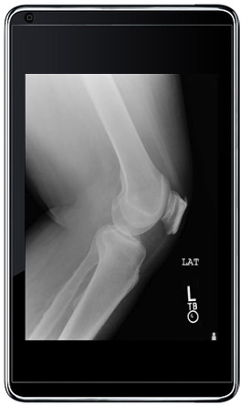 “The doctor took X-rays of my knee, sent them to XR-EXpress, where they were read by a physician in Los Angeles who then sent the report back to the XR-EXpress,” Govatski told The Hub. “The attending physician then accessed the images and radiologist’s report from his iPad and was able to show me a knee spur that could be causing the pain, and suggested I have an MRI to see if there was any soft-tissue damage.”
It is a scenario that Govatski thinks will become more and more common as the explosion of tablet devices announced at the recent Consumer Electronics Show accelerates the convergence of Internet and mobile computing.
To read the rest of this article, please visit:
It is not often a company executive has the chance to conduct a real-life test of his company’s products, but New Mexico Software CEO Richard Govatski did just that over this past holiday season. Having slipped getting into his hot tub he twisted his knee, and of course being the holiday, found himself at an urgent care clinic in Albuquerque testing the company’s new iPad app for its PACS.
The XR-EXpress iPad app allows full-function access to New Mexico Software's PACS system.
It is a scenario that Govatski thinks will become more and more common as the explosion of tablet devices announced at the recent Consumer Electronics Show accelerates the convergence of Internet and mobile computing.
To read the rest of this article, please visit:
New Mexico Software Launches Full-Function iPad App for its PACS system
Tuesday, 18 January 2011
Display Requirements for Radiology and Use of Commercial Grade Monitors
It is not uncommon for me to walk into an ER or ICU and find physicians using a commercial grade monitor that is obviously not calibrated and does not have the required contrast range needed to make a proper diagnosis. This is a major potential liability and could impact patient wellness if critical decisions are made based on viewing images of hairline fractures or a potential pneumo-thorax on these monitors.
This is not to say that one ought not use commercial grade monitors for clinical review, rather, if used for diagnostic purposes, one needs to make sure that they are properly calibrated and meet the requirements for diagnostic viewing. The question is, what are these requirements?
There are actually two sets of guidelines, one from the American College of Radiology (ACR) and one from the American Association of Physicists in Medicine (AAPM). Unfortunately, each group’s guidelines are slightly different, hard to interpret, and leave out some basic requirements such as stability and the capability for calibration and the requirement for accessing the calibration information. The latter is critical from a liability perspective.
As an example of the latter, I recently talked with a PACS administrator who was required to show the calibration records of a particular monitor that was used three years ago for a diagnosis that was later questioned in court. He had to prove that the monitor was within calibration at that time. Another example where the access to calibration data might be important is when the reading is done at a remote site, which does not have a person responsible for the image quality and integrity, so that the proper calibration can be verified and managed remotely.
To read the rest of this article, please visit:
This is not to say that one ought not use commercial grade monitors for clinical review, rather, if used for diagnostic purposes, one needs to make sure that they are properly calibrated and meet the requirements for diagnostic viewing. The question is, what are these requirements?
There are actually two sets of guidelines, one from the American College of Radiology (ACR) and one from the American Association of Physicists in Medicine (AAPM). Unfortunately, each group’s guidelines are slightly different, hard to interpret, and leave out some basic requirements such as stability and the capability for calibration and the requirement for accessing the calibration information. The latter is critical from a liability perspective.
As an example of the latter, I recently talked with a PACS administrator who was required to show the calibration records of a particular monitor that was used three years ago for a diagnosis that was later questioned in court. He had to prove that the monitor was within calibration at that time. Another example where the access to calibration data might be important is when the reading is done at a remote site, which does not have a person responsible for the image quality and integrity, so that the proper calibration can be verified and managed remotely.
To read the rest of this article, please visit:
Display Requirements for Radiology and Use of Commercial Grade Monitors
Monday, 17 January 2011
Imaging Advances And Proper Displays Are Needed For Accuracy
By Michael O'Leary:
At the center of the continuing controversy over mammograms is the potential that too frequent screening will produce too many unneeded biopsies and surgeries. As with all imaging technologies, however, mammography technology continues to advance. This issue of the Health Imaging Hub newsletter carries two stories about research advances that continue to improve the value of imaging tools for clinical practice.
The report, “Density of Mammographic Masses are a Significant Predictor of Cancer”on a research study that shows that the long suspected, but often-underutilized descriptor of suspicious lumps, may be more useful than thought. Dr. Ryan Woods and colleagues at the University of Wisconsin found that of 118 malignant lesions biopsied, 70.2 percent were high-density masses compared to 22.3 percent of the isodense or low-density masses. While it is an incremental advance, Woods points out that, “We know that mammograms are the best screening tool we have for finding cancer early, so anything we can do to improve diagnostic accuracy is useful.”
At the same time, in the article, “Advance Makes Real-time Imaging of Cellular Processes Possible” an imaging advance that may some day allow doctors to see disease processes in real time promises to improve treatments as doctors will be able to see at the cellular level whether treatments are working, or even necessary as immune cells mount a response to injury or disease.
Reporting in the journal Nature Methods, a research team led by Dr. Max Krummel, associate professor of Pathology at UCSF, has developed a new imaging technique that allows researchers to observe cellular interactions in real time without disrupting normal function. In their study they showed a series of events in an immune response to lung injury in mice. The advance overcomes the problem of blurred images produced by conventional means and results in clear images of cells interacting. A stunning series of videos are posted online in supplementary materials created by Dr. Mark R. Looney, co-first author and assistant professor in Medicine and Laboratory Medicine at UCSF.
While the clinical application of the technology is a long way off, Looney says it could soon be applied to lung biopsies. For now, Krummel says the research application could potentially lead to significant advances in understanding how cells are organized and deployed.
To read the rest of this article, please visit:
At the center of the continuing controversy over mammograms is the potential that too frequent screening will produce too many unneeded biopsies and surgeries. As with all imaging technologies, however, mammography technology continues to advance. This issue of the Health Imaging Hub newsletter carries two stories about research advances that continue to improve the value of imaging tools for clinical practice.
The report, “Density of Mammographic Masses are a Significant Predictor of Cancer”on a research study that shows that the long suspected, but often-underutilized descriptor of suspicious lumps, may be more useful than thought. Dr. Ryan Woods and colleagues at the University of Wisconsin found that of 118 malignant lesions biopsied, 70.2 percent were high-density masses compared to 22.3 percent of the isodense or low-density masses. While it is an incremental advance, Woods points out that, “We know that mammograms are the best screening tool we have for finding cancer early, so anything we can do to improve diagnostic accuracy is useful.”
At the same time, in the article, “Advance Makes Real-time Imaging of Cellular Processes Possible” an imaging advance that may some day allow doctors to see disease processes in real time promises to improve treatments as doctors will be able to see at the cellular level whether treatments are working, or even necessary as immune cells mount a response to injury or disease.
Reporting in the journal Nature Methods, a research team led by Dr. Max Krummel, associate professor of Pathology at UCSF, has developed a new imaging technique that allows researchers to observe cellular interactions in real time without disrupting normal function. In their study they showed a series of events in an immune response to lung injury in mice. The advance overcomes the problem of blurred images produced by conventional means and results in clear images of cells interacting. A stunning series of videos are posted online in supplementary materials created by Dr. Mark R. Looney, co-first author and assistant professor in Medicine and Laboratory Medicine at UCSF.
While the clinical application of the technology is a long way off, Looney says it could soon be applied to lung biopsies. For now, Krummel says the research application could potentially lead to significant advances in understanding how cells are organized and deployed.
To read the rest of this article, please visit:
Imaging Advances And Proper Displays Are Needed For Accuracy
Sunday, 16 January 2011
A New Radiology Center Opened By Papastavros
 A new diagnostic screening center has been opened recently by Papastavros' Associates in North Wilmington, North Carolina, that plays an important role in providing high –strength digital X-ray and MRI, while two other centers in Newark will provide open MRI , CT and digital X-ray. Papastavros offers detection and screening services in eight centers throughout Delaware, in addition to a number of walk-in X-ray offices.
To read the rest of the article please visit:
A New Radiology Center Opened By Papastavros
CIVCO Announces New Partnership with UltraLinq Healthcare Solutions
CIVCO Medical Solutions is announcing a co-marketing partnership with UltraLinq Healthcare Solutions of New York, New York. UltraLinq provides healthcare professionals with a secure, web-based solution for the reliable viewing, reporting, managing, sharing, distribution and archiving of multiple ultrasound and medical imaging modalities.
For more info. please visit:
For more info. please visit:
CIVCO Announces New Partnership with UltraLinq Healthcare Solutions
iSOFT’s Lorenzo Expands In Spain
iSOFT Group Limited, a leading company providing healthcare IT solutions from Australia, announced recently that is has signed a €1 million contract with Cerdanya Cross-Border Hospital Private Foundation in Spain for iSOFT’s Lorenzo Enterprise.
The Foundation is located at the border of Catalonia and France and will be the first to provide a cross-national healthcare model between Spain and France under the European Grouping for Territorial Cooperation.
According to the recently signed contract, iSOFT is going to establish an enterprise-wide clinical and hospital management system at Cerdanya Hospital, a 216-bed facility that offers elderly care, the system will be also added to seven primary care clinics in Capcir region in France, along with the Cerdanya region of Catalonia. Lorenzo Enterprise will operate all the offered clinical services. Electronic patient record (EPR) and primary care solutions of the hospital are going to use iSOFT's Viaduct integration platform in order to establish a complete enterprise-wide, clinical and patient information management system. Based on the recent contract, iSOFT is going to set up its solution at the new Cerdanya hospital, which is currently under construction, and is expected to be finished by 2013.
To read the rest of this news, please visit:
The Foundation is located at the border of Catalonia and France and will be the first to provide a cross-national healthcare model between Spain and France under the European Grouping for Territorial Cooperation.
According to the recently signed contract, iSOFT is going to establish an enterprise-wide clinical and hospital management system at Cerdanya Hospital, a 216-bed facility that offers elderly care, the system will be also added to seven primary care clinics in Capcir region in France, along with the Cerdanya region of Catalonia. Lorenzo Enterprise will operate all the offered clinical services. Electronic patient record (EPR) and primary care solutions of the hospital are going to use iSOFT's Viaduct integration platform in order to establish a complete enterprise-wide, clinical and patient information management system. Based on the recent contract, iSOFT is going to set up its solution at the new Cerdanya hospital, which is currently under construction, and is expected to be finished by 2013.
To read the rest of this news, please visit:
iSOFT’s Lorenzo Expands In Spain
Wednesday, 12 January 2011
GE And KeyHIE To Extend Healthcare IT Access In Pennsylvania
GE Healthcare, a major company providing medical imaging systems and healthcare IT solutions, and, Keystone Health Information Exchange (KeyHIE), a healthcare IT service provider in central and northeast Pennsylvania, announced that both are planning to expand the capabilities of Health Information Exchange (HIE) in the region to include chronic disease management, in addition to, improving HIE accessibility to more healthcare providers.
Jim Younkin, director, KeyHIE, said “Working with GE, we’ve achieved our initial goal of enhancing patient care by providing critical patient information, like problems, medications, and allergies to our emergency department physicians. Our next step is to use our health information exchange to accelerate collaboration among a broader set of clinicians.” Geisinger Health System, an integrated delivery network well-known for its creative use of healthcare IT care coordination, also a founding participant in KeyHIE, announced that it has received a $16 million Beacon Community award, provided by the Office of the National Coordinator for Health IT. A section of this award is going to be directed towards using KeyHIE in order to expand patient-focused care coordination throughout a community that involves around 250,000 residents.
To read the rest of this news, please visit:
Jim Younkin, director, KeyHIE, said “Working with GE, we’ve achieved our initial goal of enhancing patient care by providing critical patient information, like problems, medications, and allergies to our emergency department physicians. Our next step is to use our health information exchange to accelerate collaboration among a broader set of clinicians.” Geisinger Health System, an integrated delivery network well-known for its creative use of healthcare IT care coordination, also a founding participant in KeyHIE, announced that it has received a $16 million Beacon Community award, provided by the Office of the National Coordinator for Health IT. A section of this award is going to be directed towards using KeyHIE in order to expand patient-focused care coordination throughout a community that involves around 250,000 residents.
To read the rest of this news, please visit:
GE And KeyHIE To Extend Healthcare IT Access In Pennsylvania
MDCT Aids In Analyzing trajectories and wounds From Bullets
According to a study, published in the March issue of Radiology, Multi-detector computed tomography (MDCT) offers effective and efficient method in order to analyze wounds from bullets and explosive devices. Les R. Folio, D.O., M.P.H., the lead author of the study, from the Uniformed Services University in Bethesda, Md, mentioned that "The information provided by MDCT has the potential to improve patient care and aid in both military and civilian forensic investigations".
For more info. please visit:
For more info. please visit:
MDCT Aids In Analyzing trajectories and wounds From Bullets
Subscribe to:
Posts (Atom)